Monday, January 18, 2021

The Power of Evidence Based Uniformity In Neurologic Physical Therapy

The concept of constructing movement system diagnoses is an initiative through the Academy of Neurologic Physical Therapy.  The goal is to standardize physical therapy neurological diagnosis through consistent and evidence-based terminology describing problems we identify with the following movement systems: integumentary, neurological, musculoskeletal, cardiovascular, pulmonary, and endocrine.  The initiative is targeted to inform our examination, evaluation, diagnosis, prognosis, outcomes, and interventions.  Hedman et al. describe the need to see this initiative to fruition by stating, “while this practice may be inherent for many PTs, the profession lacks a consistent approach to movement analysis and, importantly, lacks the terminology to describe movement dysfunction in a standardized manner.”1  

In my opinion, this initiative is labeling the optimal goal that PTs and PT educators have worked toward accomplishing since the beginning of our profession.  We all want to do the best job at figuring out why our clients have difficulty moving through a detailed exam of the movement system.  We want to provide the best treatments and communicate our findings, and plan in the best way possible.  However, as our profession and vast skills evolve, we realize that we do not all agree or possess awareness relating to the optimal ways to investigate, describe, and treat these disorders.  Some of us simply lack the opportunities to train to gain the necessary skills to provide optimal care.

As a neuro/vestibular PT, I have had the opportunity to teach and mentor hundreds of therapists over the last 21 years in evaluating and treating some of the most common disorders that impact our balance system.  I have helped train these therapists onsite and through conversations over the phone, email, and texting.  I have been able to build long-term relationships with many of these therapists.  As a result, I have discovered that unless PTs are seeing a high volume of clients with dizziness and balance disorders, they will become confused when trying to complete an efficient history, objective exam, evaluation, assessment, and treatment.  

Even if PTs complete several courses directed at teaching these skills, a high caseload of clients with the appropriate diagnosis is needed consistently so that the skills can be practiced regularly.  Intense professional drive is required to put newly learned skills into practice.   If PTs do not have a high caseload of clients with the appropriate diagnosis, they will not practice the things they have learned, and concepts learned in courses can quickly be forgotten.  

Lack of patients with a key diagnosis that complements a PT's specialized training is crucial.  Without these two elements combined at the proper time in the proper environment, providing optimal quality care can be very difficult.  For instance, some PTs may forget to ask clients questions relating to the timing or triggers of symptoms.  As a result, discovering all reasons for dizziness or balance disorders may not occur.  PTs may omit core oculomotor, vestibular, and balance tests in the exam.  They may exclude postural or sensory testing.  This may lead to unnecessary discomfort for clients trying to overcome their movement disorders.  PTs may become confused when a chosen treatment is not as effective as expected, leading to an unnecessary delay in recovery.  

In summary, I support an evidence-based standardized approach to examining, evaluating, assessing (including prognosis, predicting outcomes), and providing optimal evidence-based interventions.  Building movement system diagnosis tools and databases will help provide guidance for PTs wanting to provide optimal care to all clients.  It will provide a framework that will guide decision-making, help build the best skills over larger populations of PTs, and ultimately help more people experience optimal outcomes.  

“I attest that this submission represents my own work and is compliant with Arcadia’s standards for academic integrity.”

1. Hedman LD, Quinn L, Gill-Body K, et al. White Paper: Movement System Diagnoses in Neurologic Physical Therapy. J Neurol Phys Ther. 2018;42(2):110-117.

Sunday, January 17, 2021

The Future of Outpatient Neurological Rehabilitation: Should We “Discharge” the Word Discharge?

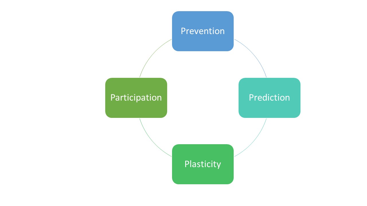
Should outpatient neurological rehabilitation therapists use the word discharge when helping clients with neurological conditions?  In my opinion, discharging outpatient clients battling neurological diseases implies an end to their journey toward being the best version of themselves they can be.  The IV STEP conference conducted in 2017 at Ohio State University provided direction for the future of neurological rehabilitation by identifying four key elements to include in patient care.1  These four themes support a holistic approach that rightfully makes discharging clients with neurological conditions in outpatient settings a treatment strategy of the past.  The four elements named include: 

  1. Prevention

  2. Prediction

  3. Plasticity

  4. Participation 

As we focus on our role as leaders in outpatient neurologic rehabilitation into the 20s and beyond, I believe these four critical themes outlined will help us expand and sharpen our focus to be involved in lifelong rehabilitation for individuals battling neurological conditions.  Lifelong rehabilitation involves helping create programs and environments.  It establishes new behaviors to help prevent the disease from getting worse. Lifelong rehabilitation encourages occasional exams through the year or years to help predict functional improvement/decline and monitor neurological performance, which helps provide accountability and leads to skilled decision making.  Lifelong rehabilitation helps guide behavioral exercises physically and mentally to optimize neuronal plasticity as our bodies change with time and includes support for participation in wellness programs long term.  These four elements are continuous with one another and really have no end.  I have included a graphic of my view of these elements in rehab of our clients below:     


We, as neuro therapists, may migrate more naturally toward one or two of the elements with our clients, but we should work hard to include all four elements in our rehabilitation plan of care.   These elements help confirm our need to be involved in our client's rehab through the entire lifespan, not just a short period of time.  I have never felt discharging clients battling neurological diseases was appropriate, and these elements support my belief.

Provide at least one example of a theme and/or key finding from the conference which you believe you already integrate into your practice regularly. 

At this time, I integrate all four elements into my practice regularly.  For instance, if I am working with a patient who has had a stroke, I will work on overall health and wellness through counseling on diet, stress management, medication compliance, sleep hygiene, aerobic and strength training, and social interaction.  I regularly take blood pressure, pulse and encourage consistent follow-up physician appointments.  All of these professional behaviors are forms of prevention.  

After working with clients who have had strokes for 21 years, I have learned how to predict which clients will recover high levels of normal movement function and which will need to learn compensatory strategies to be as mobile as possible.  I encourage plasticity through task practice and high levels of functional training daily and recommend participation in stroke support groups, exercise classes, and health and wellness programs throughout their rehab experience.  

I regularly teach my patients with neurological conditions that rehab is a lifelong process.  I do not like having to “discharge” clients from my care as I believe that terminology provides an impression that therapy is ending.  I explain to my clients that therapy will never end and that their lifelong participation in becoming the best version of themselves can be a lifelong goal.

Comment on elements from the article that surprised you, questions or concerns you have about the conference findings, AND/OR barriers you foresee in translating findings to real-world practice (either personally in your practice or as a profession). 

The comment that surprised me the most in the article was regarding the idea that certain genetic markers likely have a profound influence on motor learning and impact plasticity.1,2  Up until this point, I have usually considered non-genetic variables, such as stroke severity, medications, timing, and therapy intensity, as reasons for lack of progress following a stroke.  The idea that we could predict the likelihood for plasticity following a stroke based upon genetic makeup is fascinating.  If this type of testing and application to rehabilitation is accurate, our energy toward compensation vs. functional recovery may be impacted.

“I attest that this submission represents my own work and is compliant with Arcadia’s standards for academic integrity.”

1.Kimberley TJ, Novak I, Boyd L, Fowler E, Larsen D. Stepping Up to Rethink the Future of Rehabilitation: IV STEP Considerations and Inspirations. J Neurol Phys Ther. 2017 Jul;41 Suppl 3 Supplement, IV STEP Special Issue: S63-S72. doi: 10.1097

2.  Pearson-Fuhrhop KM, Minton B, Acevedo D, Shahbaba B, Cramer SC. Genetic variation in the human brain dopamine system influences motor learning and its modulation by L-Dopa. PLoS One. 2013;8(4):e61197.


Saturday, November 7, 2020

One of the Toughest Causes of Dizziness I See

Spontaneous spells of spinning with seconds to minutes duration can be difficult to treat. I am very interested in treatment options for vestibular paroxysmia (VP) so I wanted to conduct a literature review and see what I could find. Here is an abstract I wrote based off the following recent study:

Bayer, O., Brémová, T., Strupp, M., et al. (2018). A randomized double-blind, placebo-controlled, cross-over trial (Vestparoxy) of the treatment of vestibular paroxysmia with oxcarbazepine. Journal of neurology, 265(2), 291–298.

Purpose

Vestibular paroxysmia (VP) can be extremely frustrating to patients and clinicians because of it’s spontaneous and unpredictable nature.  Spells usually last seconds and can occur many times a month.  This monocenter, randomized, placebo-controlled, double-blind, cross-over clinical trial examined the therapeutic effect of Oxcarbazepine (OXA) in patients with VP.

Methods

43 patients between the ages of 18-80, who were diagnosed as having definite or probable VP, were enrolled for a treatment period of seven months total.  There were two treatment protocols.  Each protocol followed the same following time frames and study structure: three months of treatment or placebo followed by a one month “wash-out period” followed by the opposite placebo or treatment for three months.   OXA was the study medication and identical filling capsules were used as placebo.  Patients were randomised and evaluated by physicians periodically.  Side effects were monitored and investigators and patients were blinded to the treatment allocation sequence.

Results 

Unfortunately, the study experienced a high number of dropouts because of adverse events, relief of symptoms, or no improvement.  In the remaining participants, OXA was found to reduce the number of attacks by nearly a half (3.15 under OXA, and 5.91 under placebo treatment).

Conclusion:

OXA treatment provided significant relief compared to placebo for individuals battling VP.

Relevance to Physical Therapy

One of the greatest challenges I face as a vestibular therapist is figuring out how to stabilize spontaneous attacks of spinning lasting seconds or minutes.  In these tough cases, my role shifts from a provider of vestibular rehabilitation to a patient advocate helping triage to the most appropriate physician.  I will often encourage my clients to video their eyes during attacks to confirm the presence or absence of a vestibular problem that may be causing the attacks.  Once a vestibular problem is confirmed and characteristics fit the VP diagnosis, I can then make informed referrals to specialists.  Sometimes patients will ask what treatment options exist and I can use this study as a potential source of information.  The patient may choose to share this study with their physician which may assist in providing a more evidence based form of treatment.


Thursday, November 5, 2020

A Potential Missing Link Managing Mal De Debarquement Syndrome

Mal De Debarquement Syndrome can be extremely debilitating. I have enjoyed hearing Dr. Djalilian speak in the International Vestibular Diploma Course and wanted to dig a little deeper into some of his research. Here is an abstract I wrote based on his research on MdDS. I included some helpful links below as well.

Ghavami, Y., Haidar, Y. M., Ziai, K. N., Moshtaghi, O., Bhatt, J., Lin, H. W., & Djalilian, H. R. (2017). Management of mal de debarquement syndrome as vestibular migraines. The Laryngoscope, 127(7), 1670–1675. 

Purpose: 

Mal De Debarquement Syndrome (MdDS) is a sense of continuous movement, often described as rocking, that continues for weeks, months, or years after being on a ship, plane, train, or other prolonged continuous movement-related activity.  Unfortunately, traditional vestibular rehab can sometimes be unsuccessful at providing relief.  Nortriptyline and other migraine prophylactic medications have been reported to help suffering individuals find relief.  The purpose of this study was to investigate whether or not migraine prophylaxis would help decrease dizziness and improve quality of life in individuals suffering from MdDS.

Methods:

Clients battling dizziness and balance disorders, who presented to the researcher's tertiary neurotology clinic, were triaged into various diagnostic categories.  32 total patients met the MdDS criterion and were enrolled in their study.  15 clients were treated with education on migraine lifestyle changes and participated in the researcher's prescription-based migraine protocol.  This protocol included the following possible drugs individually or combined in various groups: Nortriptyline, Verapamil, and/or Topiramate.  Nortriptyline was the most common drug prescribed.  The trial cohort was compared with 17 past patients treated with vestibular rehabilitation and physical therapy (control group).

Results:

73% of the trial cohort had a large improvement reported using a visual analog scale (VAS).  A statistically significant difference was found in some quality of life (QOL) measures using a pre/post-treatment QOL survey.  There was minimal to no change in VAS or QOL in the control group.

Conclusion:

This study reinforces the hypothesis that MdDS will respond well to migraine prevention-based lifestyle change education combined with a special migraine prophylactic drug management protocol.  

Relevance to Physical Therapy:

Physical therapists helping clients battle MdDS should be aware that the potential cause for failure to compensate may be migrainous.  Therefore, positive treatment outcomes may be more likely when migraine prevention-based lifestyle changes such as diet, sleep hygiene, and stress reduction education are employed in conjunction with the migraine prophylactic protocol described in this study.  In my opinion, neck therapy and overall health and wellness programs should also be considered.  If improvement within three to six visits (usually over a period of two to three weeks) is not realized, a change in the treatment approach should be initiated.  It is not clear if this type of therapy was included in their control group.  


Links:

Original Research Article: https://www.ncbi.nlm.nih.gov/pmc/articles/PMC5823515/


Dr. Djalilian (one of the authors of this article) giving an outstanding lecture on Migraine management: https://www.youtube.com/watch?v=ZpK_uId5onQ&t=4750s


Tuesday, September 29, 2020

The Plastic Brain and 20/20/30

Brain imaging results and function do not always correlate with one another.  For instance, I will never forget working with a four year old many years ago.  He did not want to lie down because he was dizzy.  He demonstrated typical BPPV nystagmus, but I knew this was not likely for his age.  His positional vertigo went away in about three to four days.  He went on to have fevers and periods of ataxia that would resolve.  His speech and cognition would sometimes seem sluggish and delayed.  After months of testing, he was found to have a very rare spinal cord and brain tumor that affected his cerebral spinal fluid.  He was eventually diagnosed with a low grade glioma that is classified as a juvenile pilocytic astrocytoma. He had two cystic tumors removed- one from the cerebellum and one from the spine.  

We realized that he was amazing at compensating.  His brain was phenomenally plastic.   Imaging would reveal significant hydrocephalus with his tumors.  However, through our time in trying to figure out what was wrong, his symptoms would improve and he would “return to normal.”  We realized that his brain would compensate.  His tumors and hydrocephalus developed slowly enough that his brain would adapt.

While imaging can sometimes reveal a problem that our body can adapt to hide, sometimes our body can reveal a problem that imaging is not sensitive enough to find.  This occurs with acute brainstem strokes.  When it comes to brainstem strokes, remember 20/20/30.

I had a 45 year old gentleman who had been to the ED because of vertigo.  His CT was normal and he was told he had BPPV and referred to me.  I saw him five days later.  He described his spells as untriggered and lasting 15-30 minutes.  Spells seemed to be about two to three days apart meaning he could go a few days with no dizziness at all.  While he sat in my exam room, he said, “here it comes.”   He proceeded to have an attack of spinning and vomiting.  As I looked in his eyes, I noted direction changing nystagmus.  Since this was central sign and his history was not at all consistent with BPPV, I sent him to the ED where he progressed to have a cerebellar stroke.  He was having TIAs that the CT scans were not sensitive enough to find.  

What occurred to this gentleman was not uncommon.  20% of strokes occur in the cerebellum/brainstem and only have isolated vertigo as symptoms 20% of the time.  As a result, these kinds of strokes are missed about 30% of the time(1).   One major reason is that CT scans have 16% sensitivity (2) and diffusion weighted imaging-magnetic resonance imaging performed within 24 hours from symptom onset miss about 20% of acute posterior fossa infarctions(3).  The good news is that there is an alternative and we can help these individuals find help faster using the HINTS+ battery of  bedside tests.  A positive HINTS+ test exam of a client in Acute Vestibular Syndrome (Head impulse normal bilaterally, central appearing direction changing nystagmus, a skew deviation, and new hearing loss), is reported to suggest central pathology and have 99.9% sensitivity and 97% specificity in detecting posterior circulation infarcts(4).  I will never forget 20/20/30.

References

  1. Venhovens J, Meulstee J, Verhagen WI. Acute vestibular syndrome: a critical review and diagnostic algorithm concerning the clinical differentiation of peripheral versus central aetiologies in the emergency department. J Neurol. 2016;263(11):2151-2157.
  2. Kattah JC, Talkad AV, Wang DZ, Hsieh YH, Newman-Toker DE. HINTS to diagnose stroke in the acute vestibular syndrome: three-step bedside oculomotor examination more sensitive than early MRI diffusion-weighted imaging. Stroke. 2009;40(11):3504-3510.
  3. Newman-Toker DE. Missed stroke in acute vertigo and dizziness: It is time for action, not debate. Ann Neurol. 2016;79(1):27-31.
  4. Newman-Toker DE, Kerber KA, Hsieh YH, et al. HINTS outperforms ABCD2 to screen for stroke in acute continuous vertigo and dizziness. Acad Emerg Med. 2013;20(10):986-996.

Sunday, September 6, 2020

Diagnostic Imaging and VNGs in the Dizzy World

I am currently taking a class on diagnostic imaging.  I am going to post my hypothetical response to a patient for our discussion group here, but before I do, I want to underscore the value of a few key elements.  

#1.  When it comes to diagnosing dizziness, I want to emphasize the big three.  When it comes to helping clients figure out why they are dizzy, I always say "The Big Three is Key."  The big three are triggers, timing, and nystagmus.  Add in whether or not hearing loss is present, and we are really moving in the right direction for helping confirm the cause of dizziness.  Never underestimate the value of a high quality thorough history and bedside exam.  Also, if a client is having spells that are not present at the time of the clinical visit, have them video their eyes during an attack!  So much valuable information can be gained by doing so!  

Here is a link to the American College of Radiology's recommendations regarding when to order imaging for people who have sudden onset of dizziness and/or hearing loss.  https://acsearch.acr.org/docs/69488/Narrative/

#2.  "Normal" VNGs completed on individuals who are asymptomatic only provide insight regarding performance of the inner ear and brain at the time the VNG was performed.  Using a cell phone to record eye movements during spells should be encouraged when VNG results do not provide answers regarding why a client is dizzy.  Caution should be exercised before telling clients they have no inner ear or brain problems simply because the VNG was "normal" in the absence of symptoms.

#3.  Abnormal results on MRIs may or may not have meaning.  Please see my hypothetical response below addressed to an individual with low back pain:

You are seeing a 49 yo patient with a 4 week history of low back pain and they are very upset because their physician did not order an MRI.  Outline a potential response to this patient.  Can you imagine a scenario where the patient's frustration is justified?


I understand your concern for wanting an MRI.  However, according to the American College of Radiology guidelines, an MRI is not warranted right now because you have no red flags.  If you had a traumatic onset, a history of spinal surgery, osteoporosis, a history of cancer, suspicion of cancer, infection,  weakness or changes in your bowel/bladder, your Physician would probably be more likely to order an MRI and your frustration would certainly warrant a phone call from myself to your Physician.  These recommendations are based upon studies of thousands of different individuals combined with collaboration of experts with years of experience.  Also, please consider a study done in 2015.  The study was done on 1211 asymptomatic subjects.  87.5% had significant disc bulging, but no symptoms!(1).  Another study on 67 individuals found about ⅓ of asymptomatic individuals to have abnormal findings (2).  These individuals were surveyed over a seven year period and the abnormal findings were unable to predict which individuals would develop pain (3).  What matters most is our clinical findings at this time.   For now, I believe we can help your back pain with therapy.    

1.    Nakashima H, Yukawa Y, Suda K, Yamagata M, Ueta T, Kato F. Abnormal findings on magnetic resonance images of the cervical spines in 1211 asymptomatic subjects. Spine (Phila Pa 1976). 2015;40(6):392-398.

2.    Boden SD, Davis DO, Dina TS, Patronas NJ, Wiesel SW. Abnormal magnetic-resonance scans of the lumbar spine in asymptomatic subjects. A prospective investigation. J Bone Joint Surg Am 1990;72:403- 8

3.    Borenstein DG, O'Mara JW, Jr., Boden SD, Lauerman WC, Jacobson A, Platenberg C et al. The value of magnetic resonance imaging of the lumbar spine to predict low-back pain in asymptomatic subjects : a seven-year follow-up study. J Bone Joint Surg Am 2001;83-A:1306-11.


Thursday, September 3, 2020

Finding Answers Sometimes Takes Time...Especially if We Forget Our Roots

Reading the articles this week and watching the videos for class have reminded me of a very meaningful discussion I had with a referring ENT who had been practicing medicine 50 years.  During our conversation, I asked him what had changed the most during his time since his practice began in the 1950s.  He shared two major patterns he had identified. 

First, he stated that MRIs and CT scans had created a belief in our culture that many people could and should be diagnosed very quickly.  He explained that many people do not understand that finding a diagnosis to their problem sometimes takes time for the symptoms and disease to evolve.  He stated MRIs and CT scans had created unrealistic expectations from our patients and had also changed practice patterns of many healthcare providers.  Interestingly, Chou et al shared how these expectations can sometimes cause lack of trust in their healthcare providers if imaging is not performed. (1)  

Another great lesson my ENT friend shared is that many healthcare providers had lost the art of taking a strong history and performing a thorough bedside exam.  He stated some providers had placed too much confidence in imaging.  He stated that too many were ordering special tests instead of listening to their patients and holding true to the roots of medicine.  He explained that many had abandoned a thorough bedside exam in exchange for referring patients for more expensive testing.  

I have reflected on his observations many times over the years.  My heart breaks when I hear my clients detail the months of extensive tests and referrals they have endured.  In the context of back pain, Chou et al claim that, “routine imaging does not seem to improve clinical outcomes and exposes patients to unnecessary harms.  Imaging can lead to additional tests, follow-up, and referrals and may result in an invasive procedure of limited or questionable benefit.” (1)   

At times, I have found the dizzy world to be similar.   Many patients demand CT scans or MRIs.  They spend weeks, months, and even years going from test to test and specialist to specialist.  I have patients with dizziness spend thousands of dollars on CT scans, MRIs, VNGs, etc.  They see specialist after specialist and take months to find answers regarding why they are dizzy.  By the time they see me, they are so frustrated and have lost hope.  They lack hope in healthcare and they lack hope they will improve. In addition, they may often be in the vicious cycle of dizziness that involves avoidance, disuse, and fear that makes their problems worse.   I believe we, as passionate and knowledgeable healthcare providers, can change this paradigm through teamwork.  

Teams of healthcare providers sharing the same high quality education about the reason for symptoms followed by ways to address those symptoms and improve quality of life helps greatly.  My best results are obtained when the referring Physician and I are speaking the same language regarding the diagnosis and plan to improve.  Physicians who prepare patients for success by completing a thorough history and exam followed by a confident referral my way has lead to great outcomes.  I am often able to complete basic bedside tests and a highly sensitive/specific history that helps.  Spending time with the patient and learning how to speak their language provides them with the aha moment they are seeking.  I can often see the stress relieved from their shoulders as explanations are given through providing high quality dizziness neuroscience education.  

It is not  unreasonable for our patients to expect a quick and accurate diagnosis.  However, somehow we have to shift their confidence away from machine based diagnostics toward trusting the expertise of their healthcare provider(s).   Machine based results are often misleading.  For instance, “most lumbar imaging abnormalities are common in persons without low back pain and are only loosely associated with back symptoms.” (1)  

The bottom line is that people want to know why they are in pain or why they are dizzy.   People in general love machines and they trust those kinds of results.  However, these machines are often not able to provide the proper answers our clients seek.   We have to use our bedside exam skills, research articles available, and take the time required to provide high quality education in a way they can understand, believe, and then change their lives by changing their perspective.

1. Chou R, Qaseem A, Owens DK, Shekelle P; Clinical Guidelines Committee of the American College of Physicians. Diagnostic imaging for low back pain: advice for high-value health care from the American College of Physicians [published correction appears in Ann Intern Med. 2012 Jan 3;156(1 Pt 1):71]. Ann Intern Med. 2011;154(3):181-189.